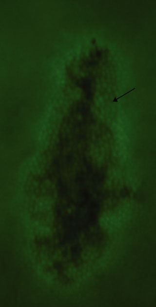
Figure F280. Epifluorescent image of cells grown on a marine agar 2216 plate (sample from Section 305-U1309D-80R-1) (field of view = 30 × 60 µm). Cells stained with acridine orange appear as light green spheres. Arrow indicates a cluster of cells.
Previous | Close | Next | Top of page